Fiber-optic Two-Photon Endomicroscope for Functional Histological Imaging In Vivo
Speaker
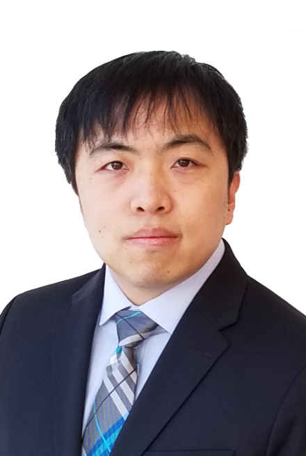
Wenxuan Liang

Time
2020-01-02 10:00:00 ~ 2020-01-02 11:30:00
Location
Room 1-402A, SEIEE Building
Host
YuYe Ling, Assistant Professor, John Hopcroft Center for Computer Science
Abstract
Optical microscopy is indispensable for conventional histopathological analysis of biopsy specimens. The entire procedure, although regarded as the gold standard for diagnosis, is well-known to be prone to sampling error, time-consuming (a few days typically); and particularly, unable to reveal any in vivo functional information. Two-photon microscopy (TPM), with intrinsic advantages in penetration depth and three-dimensional resolution, can be a perfect candidate for in vivo histology. However, clinical translation of TPM has been hampered by the bulky footprint of standard bench-top TPM platforms. Therefore, a miniature two-photon endomicroscope has been long envisaged to furnish direct imaging of internal biological tissues in vivo. Such an enabling compact imaging platform can also fulfill the need in a variety of basic research applications, such as brain imaging on freely-behaving animals. Our lab has pioneered the development of a fiber-optic, miniature two-photon endomicroscope of an ultralight weight (<1 gram) and an ultracompact size (~2 mm in diameter). In this presentation, I will discuss the basic design concept of the technology and the recent breakthrough we have achieved to significantly improve the imaging signal-to-noise ratio (i.e. by 20-50X compared with the prior art), which enables label-free imaging of internal organs in vivo, in situ, and in real time, with the imaging quality comparable to the standard bench-top counterpart. I will also present the preliminary results of this technology in a variety of potential applications, including assessment of (1) preterm-birth risk by visualizing cervical collagen fiber network and (2) cellular metabolic function (redox ratio) based on the intensity and lifetime of NADH autofluorescence imaging on live animal models. Initial results of two-photon imaging of spontaneous responses of astrocytes on freely-walking mice will also be presented.
Bio
Wenxuan Liang earned his bachelor’s degree and Master’s degree, both in Biomedical Engineering (BME), from Tsinghua University, and then completed PhD degree in BME from Johns Hopkins University School of Medicine. His PhD thesis focuses on developing a flexible fiber-optic miniature two-photon endomicroscope, which achieves unprecedented detection sensitivity and therefore enables label-free imaging of internal organs in vivo, in situ, and in real time. The miniature fiberscope opens up numerous new opportunities, from label-free histological visualization of internal organs for early disease diagnosis and biopsy guidance, to brain imaging on freely-behaving animals. He is currently working at Columbia University as a postdoctoral scientist, developing a next-generation single objective ultrafast 3D light-sheet microscopy technology, named swept confocally-aligned planar excitation (SCAPE) microscopy, which enables real-time calcium imaging of thousands of neurons across multiple cortical layers from awake, behaving mammals, and brain-wide neuronal network dynamics of small organisms including fruit flies, zebrafish, and C. elegans worms.

